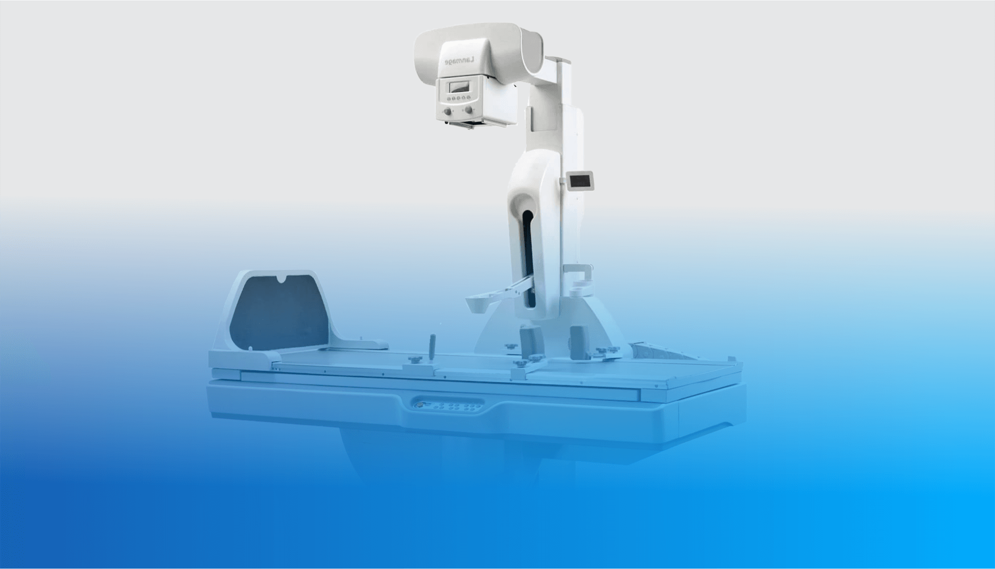
Fluoroscopy
What Is Fluoroscopy?
Fluoroscopy is a medical imaging technique that uses X-rays to produce real-time images of the inside of a patient's body. The procedure involves the use of a fluoroscope, which is a specialized X-ray machine that produces a continuous stream of X-ray images that are displayed on a monitor.
During a fluoroscopy procedure, the patient is typically positioned on a table or in a chair, and a contrast agent may be administered to help highlight specific areas of the body. The X-ray machine emits a continuous beam of radiation that passes through the patient's body, and the resulting images are captured and displayed on a monitor in real-time.
Some Of The Advantages Of Fluoroscopy include:
-
Visualization: Fluoroscopy allows doctors to see live images of internal structures, which can be useful in guiding procedures and ensuring accurate placement of medical devices.
-
Non-invasive: Fluoroscopy is a non-invasive procedure that does not require surgery or incisions. This can be beneficial for patients who are not good candidates for more invasive procedures.
-
Low radiation dose: With modern technology, fluoroscopy can be performed with a lower radiation dose than in the past, reducing the risk of radiation exposure for patients and medical personnel.
-
Versatility: Fluoroscopy can be used for a variety of procedures, including diagnostic tests, biopsies, and interventional procedures such as placing a catheter or pacemaker.
-
Cost-effective: Fluoroscopy is generally less expensive than other imaging techniques such as CT or MRI scans, making it a more cost-effective option for certain procedures.
-
Wide availability: Fluoroscopy equipment is widely available in most hospitals and clinics, making it accessible for patients who need the procedure.
"Fluoroscopy is a key technology in interventional radiology, enabling us to treat a wide range of conditions with minimally invasive techniques." - Dr. Michael D. Dake

